
The movements involved in ice skating put a lot of pressure on the knee joint. When you combine those stressful movements with a highly physical, collision-filled sport like hockey, the knee becomes one of the sites most susceptible to injury. Injuries to the three main ligaments in the knee – the anterior cruciate ligament (ACL), medial collateral ligament (MCL) and posterior cruciate ligament (PCL) – are all relatively common in hockey, with ACL and MCL the more frequent.
Connor Mc David – a double Ted Lindsay Award winner and one of the fastest skaters in hockey – damaged his knee by colliding with the goal in the last game of the 2018-19 season. X-rays initially suggested the impact had caused no serious damage, but a precautionary MRI scan proved otherwise. A small tear was found in McDavid’s posterior cruciate ligament and at his next public appearance he was wearing a knee brace. Read more about McDavid’s experience here.
Particularly in cases of high-speed impact, x-ray is often the first method used to examine an injured knee, due to the high chance of bone fractures. However, x-ray cannot show ligament injuries, so if nothing shows up but there is still a strong suspicion of damage, MRI is a common next step, especially for professional athletes.
MRI is the best radiological tool for evaluation of the entire joint because on MR images it is possible to see deeply located anatomical structure like cruciate ligaments. This MR image, in the sagittal plane, shows incomplete disruption of the posterior cruciate ligament fibres (arrow).
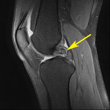
Note: image is an example – not that of the athlete named above.
MRI is the method of choice for showing small tears in ligaments and can provide a detailed visualisation of the complex structure of the knee. The radiologist knows exactly how to interpret the images and assess the full extent and location of the damage – crucial information that will help the athlete’s medical team to plan recovery.
For more information about PCL tears, click here.