
Anyone who has tried skiing knows exactly how much it depends on the knees. Constant pressure is placed on the ligaments of the knee joint as the leg muscles react to dynamic forces in every direction. So, it will come as no surprise that knee injuries – especially to those ligaments – are among the most common in skiing.
In January 2019, Olympic silver medallist and former slalom Junior World Champion, Stephanie Brunner, hurt her knee during a training session. The knee was examined with MRI, which revealed damage to her meniscus (the shock-absorbing cartilage in the centre of the knee) as well as a torn anterior cruciate ligament – the most frequently occurring injury by far among World Cup skiers. Read more about Brunner’s injury here.
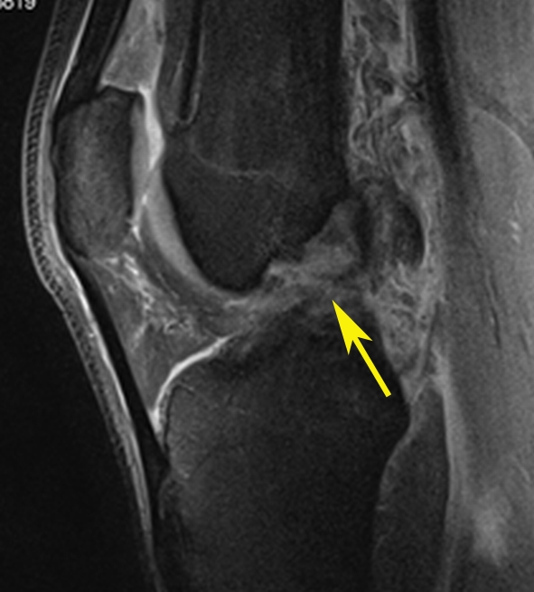
MRI is the best radiological method for evaluating the entire joint, especially in cases of injuries of multiple anatomical structures of the knee. The MR images below show a case of complex injury of three important knee structures.
The sagittal view shows complete disruption of the anterior cruciate ligament (arrow)
The coronal view shows a lateral meniscus tear (yellow arrow) and complete disruption of the medial collateral ligament fibres (red arrow)
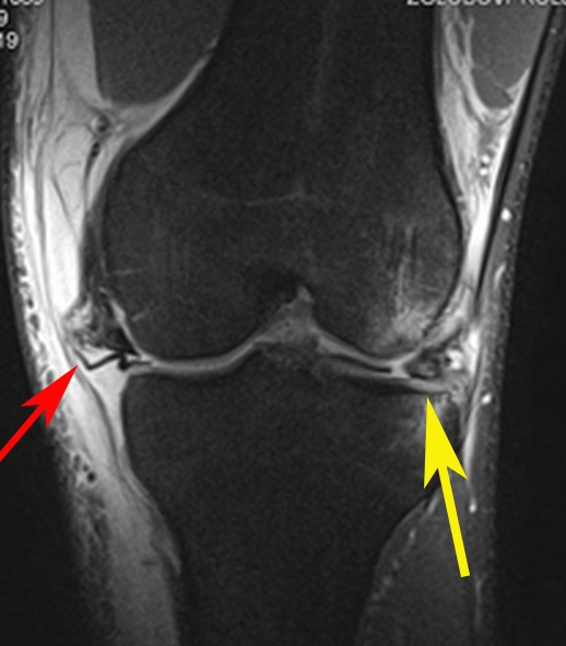
Note: images are examples – not those of the athlete named above.
One of the most basic principles of radiology is to avoid ‘satisfaction of search’. This means that when the radiologist is analysing an image and finds the likely cause of the problem, they are never satisfied that the search is over! As in Stephanie Brunner’s case, it is often possible that there is more than one area of damage, so radiologists are always sure to conduct the most thorough investigation possible, in order to help the athlete to plan recovery and return to action as quickly as possible.
For more information about knee injuries in skiing and snowboarding, click here.