Back injuries are very common among tennis players. Constant rotation of the trunk and spine, as well as stop-start motions, put a lot of stress on the back muscles. Hyperextension of the lower back during serving can also cause cumulative damage to the vertebrae, ligaments and tendons, as well as compression of the lumbar discs.
Former world no.1 Simona Halep was forced to pull out of her first-round match at the 2018 China Open due to the return of a previous back injury. An MRI examination two days later revealed that the four-time Grand Slam winner had a herniated disc. Read more about her experience here.
Small discs of cartilage act as cushions between the individual vertebrae. Each disc consists of a tough outer ring and a softer nucleus. Excessive wear and pressure on the disc can cause part of the softer nucleus to be forced through a tear in a damaged outer ring, into the spinal canal. This can put pressure on spinal nerves, causing considerable pain.
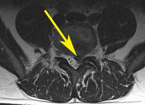
MRI is an essential diagnostic method for assessing herniated discs. This MR image of the lumbar spine in the transverse plane shows a herniated intervertebral disc. The bulging part of the disc is pushing into the space where the neural root passes through (arrow).

Note: image is an example – not that of the athlete named above.
MRI is just one of many examinations that may potentially be used to diagnose a suspected herniated disc and radiologists are the experts who analyse the resulting images. The examining radiologist will determine nature of the injury, the exact location and possible nerve root compression, and will be able to spot any associated complications such as an impacted nerve.
For more information about herniated discs, click here.
