Knee injuries are very common at all levels of football, with ligament damage accounting for a high percentage. The anterior cruciate ligament (ACL) is one that makes particularly regular appearances in football injury reports. The ACL is one of two ligaments that form a ‘cross’ shape behind the kneecap, connecting the thighbone to the shinbone. The frequent stopping, starting, twisting and turning required of footballers puts a large amount of strain on these ligaments and can lead to strains, tears and even complete ruptures.
US soccer star Megan Rapinoe suffered her own ACL tear in December 2015, damaging her right knee while training with the national team. The injury – which was the third ACL tear of Rapinoe’s career – was confirmed with an MRI examination. Ultimately, she recovered in time to take part in the 2016 Olympics. Read more here.
MRI is the best radiological tool for evaluation of the entire knee because it is possible to see the anatomical structures that are located deep within the joint.
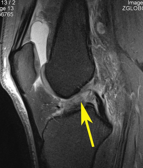
A typical ACL tear looks something like this:

Note: image is an example – not that of the athlete named above.
To a radiologist, this MR image clearly shows the complete disruption of the anterior cruciate ligament fibres (arrow). By examining images like this one, expert radiologists can identify the location and extent of damage with great accuracy and provide valuable information that helps treatment and recovery.
For more information about ACL injuries, click here.
