
Cricket is played with a relatively hard ball compared to most other sports and the players making the catches in the field do not wear gloves. Naturally, this means finger injuries are an occupational hazard for cricketers, with sprains, dislocations and fractures common to the game.
Eoin Morgan, captain of the 2019 World Cup winning England cricket team, suffered a fracture and dislocation less than a week before that tournament began. Morgan’s left index finger was struck with a ball during catching practice. He was immediately sent for x-rays, which showed a “small flake fracture” (whereby a small flake is chipped from the bone’s surface) in addition to the obvious dislocation. The diagnosis led to him missing just one warm-up game before re-taking his place in the team for the start of the World Cup. Read more about Morgan’s injury scare here.
Finger dislocations are easy to spot with a physical examination, but such an injury should always be checked with x-ray to determine the presence of any fractures. X-ray is the first choice for bone-related investigations as it shows the contrast between bone and soft tissue extremely well. When a radiologist examines images of a dislocated finger, they know the locations to check for the most likely possible fractures and can spot even the smallest chips and cracks in bone.
X-ray is the best radiological method for detecting bone fractures and the positions of fractured fragments. The arrow in the image below indicates the location of a ‘medial phalanx avulsion fracture’. The medial phalanx is the middle bone of the finger and an avulsion fracture occurs when trauma causes a small fragment of bone to be pulled away from the main bone. In this case there are no dislocated fragments.
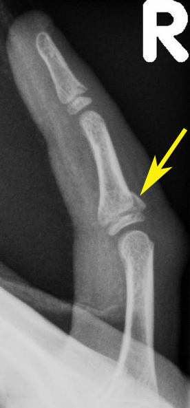
Note: image is an example – not that of the athlete named above.
For more information about finger fractures click here.