
In a sport that involves swinging a club with great force, it is no surprise that a lot of golfers suffer wrist injuries. Bones, muscles, tendons, nerves and even blood vessels can be damaged by repetitive impact or simple overuse. One such typical injury is nerve entrapment or nerve compression syndrome, which can often be a secondary result of an inflammation elsewhere.
In March 2019, former US Women’s Open winner, Michelle Wie, was forced to withdraw from the Women’s World Championship in Singapore after experiencing so much pain that she was “unable to lift the club”. MRI images later revealed that she was suffering from nerve entrapment due to inflamed tendons. In other words, the swelling of the tendons was irritating the adjacent nerve so much that it caused her intense discomfort. Read more about Wie’s experience here.
Nerve compression will usually be diagnosed by physically examining and talking to the patient, but medical imaging is essential in order to find the underlying cause. When radiologists want more information about a whole joint or anatomical region, such as to detect the cause of nerve compression, MRI is method of choice. Below is an example of such a post-traumatic nerve compression shown on MR images. Images in the coronal plane (image 1) and axial plane (image 2) clearly show fluid (arrows) in the tendon sheets, which is compressing the median nerve (reactive tenosynovitis).
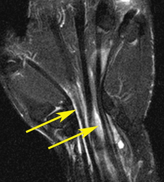

Note: images are examples – not those of the athlete named above.
Radiology staff, including sonographers, radiographers and radiologists are all crucial to the process, combining their skills and training to gather the most useful images, interpret them and pass on a detailed report.
For more information about nerve compression, click here.