Big powerful players, lots of running, and full physical contact are all factors that make rugby a hotbed of sports injuries, especially those involving the knee. The pressure put on rugby players’ knees results in a lot of soft tissue complaints, such as sprains and tears of the ligaments that hold the knee joint together.
Australian international David Pocock suffered a knee injury in 2013 while playing for his club side, the Brumbies. After a tackle by an opposition player, Pocock had to be helped from the field and was later examined with MRI. The resulting images showed that he had torn his anterior cruciate ligament (ACL), one of two small ligaments that form a cross shape in the centre of the knee joint. Read more about Pocock’s case here.
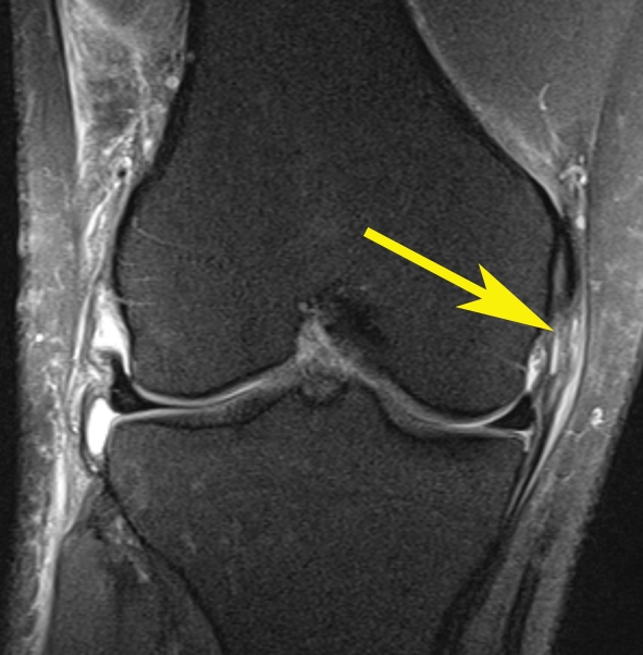
Although ultrasound is the simplest method for superficial ligament evaluation, MRI is the best radiological method for evaluation of the entire joint, especially in cases of injuries of multiple anatomical structures of the knee. The MR image below, in the coronal plane, shows the subtle signs of incomplete disruption of the medial collateral ligament fibres (arrow).

Note: image is an example – not that of the athlete named above.
ACL injuries are an extremely common occurrence in many sports. It is possible to diagnose ACL damage with a physical examination, but an imaging examination such as MRI is always needed to assess the exact location and full extent of the injury, and to check for damage to any other tissues. The radiologist is the expert who makes the assessment, based on a detailed knowledge of anatomy and how it should – and shouldn’t – look on images.
For more information about soft tissue knee injuries in rugby, click here.
