
Spinal injuries comprise a relatively small proportion of athletic injuries, but they are nevertheless a realistic risk in many sports. Some sports carry an increased risk due to the nature of the repetitive motions involved or the frequency of contact with the ground, water, or other athletes. Divers, for example, have been observed to sustain a particularly high rate of spinal injuries1.
Australian diver Taneka Kovchenko was forced to retire from the sport in 2017 after medical images revealed that two of her vertebrae were putting dangerous pressure on her spinal cord. Kovchenko’s initial scans, which were carried out to find the cause of her head and neck pain, did not show anything suspicious. But, as she notes in an Instagram post, she later had additional scans with her neck in other positions, which showed that when she tipped her head back, the C1 and C2 vertebrae were compressing her spinal cord. You can read more about Kovchenko’s experience here.
As in Kovchenko’s case, radiologists – and the radiographers who actually obtain the images – know that the root cause of a problem might only be spotted with the head or neck in a particular position. This kind of expert knowledge can literally save lives.
Although MRI is often the first choice for examining soft tissues, CT enables visual reconstructions of the body with multiple views and in 3D. This is especially useful for detecting the positions of fracture fragments that may put pressure on other structures. The sagittal reconstruction CT image below clearly shows a fragment of dislocated bone (arrow) putting pressure on the spinal cord and nerve roots.
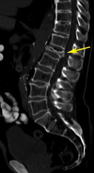
Note: image is an example – not that of the athlete named above.
For more information about spinal compression injuries, click here.